WHAT IS COMBINED FIRST TRIMESTER SCREENING?
Combined first trimester screening involves the markers of the ultrasound (popularly called as NT, NB scan) and the blood test- dual marker test. These markers on the scan and the values of the blood test together when combined, provides a risk assessment which tells us that the pregnancy is at high risk or not for genetic abnormalities like Trisomy 21 (Downs syndrome). For risk assessment, we use a software provided by Fetal Medicine Foundation, UK.
WHAT IS THE PURPOSE OF THE SCAN?
The aim is to establish that the baby is alive or not and correctly, establish its gestational age. It involves assessing the baby for gross structural abnormalities. The most important aspect is to assess certain markers like fluid behind the neck of the baby (nuchal translucency), presence of nasal bone, blood flows through heart (tricuspid Doppler) and tummy (ductus venosus) and heart rate. These markers on the scan and the values of the blood test together provide a risk assessment which tell us that the pregnancy is at high risk for chromosomal abnormalities like Trisomy 21 (Downs syndrome). For risk assessment, we use a software provided by Fetal Medicine Foundation, UK. It also provides us an opportunity to assess the pregnancies who could be at high risk for developing high blood pressure or weak baby during the course of the pregnancy.
WHAT PROTOCOL IS FOLLOWED WHILE DOING SCAN?
We perform the scan in accordance with the guidelines and the protocol laid down by ISUOG (International Society of Ultrasound in Obstetrics and Gynaecology) and FMF (Fetal Medicine Foundation, UK).
WHEN IT IS DONE?
It is done between 11-13+6 weeks.
WHO SHOULD GET IT DONE?
Ideally, all the pregnant females should be offered combined first trimester screening.
WHO SHOULD DO IT?
Individuals who perform obstetric scans routinely should have specialized training that is appropriate to the practice of diagnostic ultrasound for pregnant women. We have FMF, UK certificate of competence for NT, NB scan. Our work is audited every year so that we maintain the quality standards as laid down by international organizations. Besides the scan, we will also guide you what is to be done next if a particular abnormality has been detected on the scan.
IS PRENATAL ULTRASONOGRAPHY SAFE?
Prenatal ultrasonography appears to be safe for clinical practice. To date, there has been no independently confirmed study to suggest otherwise.
WHAT IS CERVICAL LENGTH SCREENING?

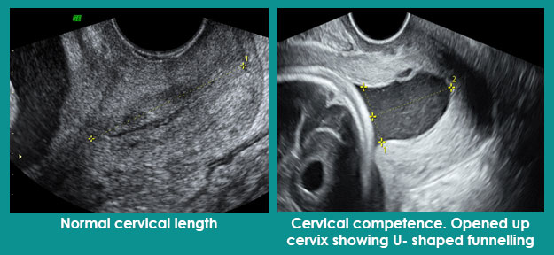
The cervix is the lower part of the uterus (mouth of the uterus). It is this that shortens and opens up allowing the passage of baby during the childbirth. We want it to remain long and closed throughout the pregnancy and open up only at the time of childbirth.

Measuring the length of the cervix is called as cervical length screening.

WHAT IS THE PURPOSE OF THE SCAN FOR CERVICAL LENGTH SCREENING?

The purpose is to determine whether the cervix is short or long and whether it has started opening (called as funnelling) up or not. Any cervical length which is < 25 mm is labelled as short cervix.

Pregnancies with short cervices are at increased risk of second trimester miscarriage or delivery before 34 weeks.

HOW THE SCAN FOR CERVICAL LENGTH MEASUREMENT IS DONE?

It is always done vaginally. We can do it from abdomen as well, however, that assessment for length is spurious.

WHEN IT IS DONE?

There is no specific time period. We do cervical length measurement transvaginally at the time of first trimester screening as it provides us a baseline. Then depending on women’s previous history of preterm delivery or second trimester miscarriage or clinical complaints like pain or heaviness in the lower abdomen, bleeding or leakage of fluid per vaginum, we do an internal scan for cervical length assessment.

WHAT PROTOCOL IS FOLLOWED FOR CERVICAL LENGTH MEASUREMENT?

We follow the protocol as laid down by Fetal Medicine Foundation (FMF), UK.

IS IT SAFE TO DO A TRANSVAGINAL ULTRASOUND?

This is the most common question we are asked. Transvaginal ultrasound is absolutely safe. It will not harm the baby or induce miscarriage or bleeding. At the most, you might feel a slight discomfort while undergoing transvaginal scan but then this also depends on your pain threshold.

Cervical Length Screening
WHAT IS FETAL ECHOCARDIOGRAPHY?
Fetal echocardiography is nothing but a detailed ultrasound the fetal heart. The heart and its connections, blood flows and their velocities are checked to identify and characterize the fetal heart anomalies
IS FETAL ECHOCARDIOGRAPHY DIFFERENT FROM THE ANOMALY SCAN?
Fetal echocardiography is only about fetal heart evaluation whereas in an anomaly scan, besides heart, other structures are also assessed. Normally, fetal heart assessment requires minimum evaluation of 4 chamber view, outflow tracts and 3 vessel views.
Many people restrict the assessment to 4 chamber view and outflow tracts and some only to 4 chamber view. This is not enough to rule out cardiac abnormalities. If these are not assessed in the anomaly scan, then fetal echocardiography is advised. We assess the fetal heart in detail in anomaly scan only.
WHO SHOULD UNDERGO FETAL ECHOCARDIOGRAPHY?
Fetal echocardiography is indicated if there is suspicion of cardiac structural or functional abnormality, abnormal fetal heart rate or rhythm, multiple pregnancy, increased nuchal translucency (fluid behind neck of the baby), extra-cardiac abnormality or if mother has diabetes or auto-immune disease, history of heart disease in parents, siblings or previous pregnancy. Obesity or suboptimal views or suboptimal assessment during anomaly scan can also warrant fetal echocardiography.
CAN WE DETECT ALL FETAL HEART ABNORMALITIES ON FETAL ECHOCARDIOGRAPHY?
No, we cannot. There are certain lesions like aortic stenosis, coarctation of aorta which might get apparent later as they evolve over time. However, the aim is to maximise the probability of detecting and correctly identifying clinically relevant congenital heart diseases.
WHEN IS IT DONE?
It is usually done between 18-22 weeks, although some structure might be better identified before or after this period. It is possible to assess the fetal heart early like at the time of first trimester screening scan when certain heart defects can be recognised or atleast a suspicion can be raised.
WHAT PROTOCOL IS FOLLOWED FOR DOING FETAL ECHOCARDIOGRAPHY?
We follow the protocol as laid down by ISUOG (International Society of Ultrasound in Obstetrics and Gynaecology) 2013 and AIUM (American Institute of Ultrasound Medicine) 2020.
IS ULTRASOUND SAFE DURING PREGNANCY?
Ultrasonography is absolutely safe for clinical practice. To date, there has been no independently confirmed study to suggest otherwise.
WHAT IS AMNIOCENTESIS?

Amniocentesis is a test offered in the second trimester of pregnancy. A small sample of fluid is taken from the amniotic sac using the needle. The amniotic sac is the sac which contains the baby and the fluid around it inside the uterus. This fluid around the baby has baby’s shed cells. These cells are extracted and used to check if there is a chromosomal or any other genetic problem in the baby.

WHAT IS THE PURPOSE OF THE AMNIOCENTESIS?

Amniocentesis is offered

a. If prenatal screening shows there is an increased chance of a chromosomal problem like Down syndrome,
b. If an ultrasound scan identifies a structural problem in the baby that requires further testing, or
c. If there is a family history of a chromosomal problem or a known genetic condition.

Getting the test done is completely your choice. We can suggest what is good practice based on evidence-based medicine. Knowing all the pros and cons, you can accept or decline the testing.

WHEN IT IS DONE?

It is usually done after 15 completed weeks. It can be done uptil any time, however, in the third trimester the number of cells in the fluid decreases, so might not be able to give any result. We usually do it between 16 to 20-24 weeks. The MTP act allows termination till 20 weeks, hence we prefer, it to be done earlier than 20 weeks.

HOW IT IS DONE?
Amniocentesis is done by fetal medicine specialists and will take a few minutes only. You do not need a full bladder. First, a scan is done to look at the position of the baby and the placenta. This helps to guide the needle into the womb. The fluid should be clear and yellow. It is baby’s urine which will get naturally replaced in a day or two.

What you feel is only a prick as the needle goes through the skin into the fluid around the baby. A little pressure is felt as the fluid is withdrawn into the syringe. So, most women feel slight discomfort but no pain.
WHAT ARE THE RISKS INVOLVED?

There is minimal risk of miscarriage about 1: 1000. We do not know why it happens or to whom it will happen. It does not cause any birth defects or any harm to the mother. The risk of infection is also very low, 1 in 1000.

WHAT HAPPENS AFTER THE PROCEDURE?

You are made to rest for half an hour after the procedure. The fetal heart beat is checked and then you are sent home. Most women have no problems, but you might have a bruise, spotting or cramping. If you do, paracetamol is safe to take. If you have a persistent pain, leaking, bleeding or a high temperature, then you need to go to the hospital.

WHOM TO CONSULT?

You can consult with Dr. Neha Gupta. She is one of the best fetal medicine specialists and is experienced in handling lots of pregnant women to take care of their unborn baby. Contact us to get the appointment and discuss it with her.

WHAT IS MULTIFETAL PREGNANCY REDUCTION?
Multifetal pregnancy reduction is a procedure that involves reducing the total number of babies in a multiple pregnancy by one or two.
WHAT IS THE PURPOSE OF MULTIFETAL PREGNANCY REDUCTION?
The cases of multiple pregnancy are increasing around the globe especially because of the advanced maternal age at pregnancy and more and more use of artificial reproductive techniques. When there are more than one fetuses in your womb, there is a possibility of miscarrying the entire pregnancy or early delivery. Babies who are born too soon have the risk of developing jaundice, infection, injuries to brain, breathing problems, feeding problems and require ICU care.
Mothers are also at increased risk of complications during pregnancy like high blood pressure, high sugars, anaemia, preterm delivery. These problems are more pronounced especially when there are 3 or more babies. Hence, your obstetrician will suggest you to consult a fetal medicine specialist to discuss about multifetal pregnancy reduction. Multifetal pregnancy reduction is also offered in those cases in which one of the babies has a lethal structural abnormality
WHEN IT IS DONE?
The procedure is done in the first trimester or early second trimester. It is generally done between 11-12 weeks.
HOW IT IS DONE?
It’s usually done per abdomen. First, an ultrasound is done to know the type of multiple pregnancy. We look for structural abnormality in any of the babies in your womb. If the initial screening looks ok, we select the baby that is far away from the mouth of uterus and is easily accessible. With the help of ultrasound, a local anaesthetic is injected into the underlying skin so that you don’t feel any pain. We introduce the needle from the tummy into the selected fetus’s heart. We inject the medicine like KCL into the heart. The heart beat stops. We observe for a little while and then the needle is withdrawn. Those multiple pregnancies where the placenta is shared, a different procedure called laser or radiofrequency ablation is used. The fetal medicine specialist will discuss with you if this kind of procedure is required.
IS IT SAFE?
Usually, it is a safe procedure. However, there is minor risk of miscarrying the entire pregnancy. What we have to consider here is the risk benefit ratio analysis. Carrying a multiple pregnancy, increase the risk of preterm delivery by 60% and that to too early. This procedure reduced the risk by half. The fetal medicine specialist will discuss in detail with you.  
WHOM TO CONSULT?
Our one of the best fetal medicine consultants Dr. Neha Gupta specializes in this area. You can consult with her. You can call us and request an appointment.
WHAT IS IUT??

An Intrauterine transfusion (IUT) is a procedure in which blood is transfused into a fetus, most commonly through the umbilical cord. It is used in cases of severe fetal anaemia.

WHEN IT IS DONE?


Fetal anaemia occurs in haemolytic disease of the fetus and newborn.

For HDFN to occur, the fetus must have a positive blood group (paternally inherited) and the mother must have negative blood group. Fetal red cells has the Rh factor which is the antigen which the mother does not have. When mother is exposed to the Rh factor during a previous pregnancy or transfusion, mother develops antibodies to the Rh factor.

In the next pregnancy, maternal antibodies cross the placenta during pregnancy and destroy fetal red blood cells (RBCs). This process can lead to fetal anaemia, and in severe cases can progress to hydrops (edema), ascites, heart failure, and death.

However, the rates of these cases have significantly reduced by Anti-D immunoglobulin administration.

Fetal anaemia is monitored throughout pregnancy using Doppler measurement of the middle cerebral artery (MCA) peak systolic velocity (PSV). Once MCA-PSV value exceeds 1.5 MoM, it indicates development of fetal anaemia. At this point, invasive testing via percutaneous umbilical cord blood sampling (PUBS, also called cordocentesis) is done followed by fetal transfusion

HOW IT IS DONE?

Prior to the procedure, the compatible blood is obtained. O, RhD-negative, and antigen-negative for maternal RBC antibodies is selected. The selected blood then undergoes irradiation and leukocyte reduction. Antenatal corticosteroids are typically given to mothers before IUT to anticipate the need for an emergency caesarean section.

The procedure is usually performed in a hospital under sterile conditions. The mother’s abdomen is cleaned with an antiseptic solution. She is given a local anaesthetic injection to numb the abdominal area from where the transfusion needle will be inserted. During the procedure, medicine is given to the fetus in its thigh to temporarily stop the fetal movements.

An ultrasound is performed to view the position of the fetus and to help guide the needle. The first step is to locate a relatively stable segment of the umbilical cord. Once a suitable location is established, the needle is inserted through the mother’s abdomen into an umbilical vessel using ultrasound guidance. If insertion into an umbilical vessel is not possible, blood may be transfused into the portion of the umbilical vein in the fetal abdomen.

Prior to the transfusion, percutaneous umbilical cord blood sampling (PUBS) is conducted. The fetal blood sample is drawn and immediately analysed for haematocrit in the hospital haematology laboratory. The result confirms the level of fetal anaemia and indicates the correct amount of blood to be transfused. With the needle still in place, the blood is delivered into the fetus’s umbilical cord blood vessel. Following the transfusion, an additional blood sample is drawn and analysed to determine the final hemoglobin.

WHAT ARE THE RISKS?

Fetal survival rates after intrauterine transfusion through the umbilical cord are more than 90% for fetuses that do not have hydrops and about 75% for fetuses that have hydrops

Risks of intrauterine transfusions may include uterine infection, fetal infection, preterm labour, excessive bleeding and mixing of fetal and maternal blood, amniotic fluid leakage from the uterus, slowing of fetal hearty rate, bleeding from the umbilical cord puncture site or (rarely) fetal death. There is only 1-2% risk of such complications.

Laser And Radiofrequency Ablation Procedures

WHAT ARE ABLATION PROCEDURES?
These are procedures in which we use additional energy like radiofrequency waves or laser to stop the blood flow in an abnormal fetus. It is delivered to the fetus by means of a needle inserted into the abnormal baby. It uses a small needle that is inserted under ultrasound guidance into the tummy of the baby at the site of umbilical cord insertion.
Usually two types of ablation procedures are there-
That Uses Radiofrequency Needle – Popularly Called As RFA Procedure. The Needle Is Then Connected To The Radiofrequency Generator. Once Turned “On”, These Waves Coagulate The Blood Vessels. It Is Done Until There Is No More Blood Flow Going To The Fetus. The Advantage Of This Technique Is The Small Size Of The RFA Needle Which Minimizes The Risks Of Preterm Labour. The Ablated Fetus Gets Reabsorbed Or Gets Reduced Toa Much Smaller Size.The Other Uses ND-YAG Laser- In This, The Laser Fibre Is Passed Through The Needle And It Is Then Connected To The Generator Producing Laser Waves. However, It Is Used At Lower Gestations When The Fetus Is Small.
WHICH CONDITIONS ARE TREATED WITH ABLATION PROCEDURES?
Ablation procedures are typically utilized in abnormal twin pregnancies. This is usually seen in MONOCHORIONIC TWIN pregnancies- one which in which the babies share their placenta.
It Is Commonly Used In Twin Reversed Arterial Perfusion Sequence (TRAP) Or More Popularly Called As Acardiac Twin.Other Conditions Where It Can Be Used To Stop Blood Flow Of One Of The Twins Is In Monochorionic Pregnancies Where There Is Structural Abnormality In One Twin.There Is Development Of TTTS – Twin To Twin Transfusion Syndrome.
The need to perform ablation procedures will be determined by the fetal medicine specialist.
WHAT IS TRAP (TWIN REVERSED ARTERIAL PERFUSION SEQUENCE)?
Twin reversed arterial perfusion (TRAP) sequence or acardiac twin is a very rare and unique problem. It is complication seen in approximately 1% of monochorionic twins (twins sharing the placenta). One twin is structurally completely normal. The other is an abnormal mass of tissue, consisting usually of legs and a lower body, but no upper body, head or heart. Because of the absence of heart, the term acardiac twin is used. The normal fetus is the “pump twin” because its heart is used to pump blood to the abnormal mass. The acardiac twin has no chance of survival. It receives and pumps back the blood supply from the normal twin via vascular connections present on the surface of the placenta. The normal twin’s heart has to additional work of pumping the blood to the abnormal one. Due to this the normal twin develops cardiac failure and if nothing is done, then eventually dies.
WHAT IS TTTS- TWIN TO TWIN TRANSFUSION SYNDROME?
This is a unique complication seen in Monochorionic pregnancies. Due to presence abnormal vascular communications on the surface of the placenta, one twin acts as pump and the other as recipient. Pump twin pumps blood to the other via these vascular connections. As a result, the pump twin goes small, develops anhydramnios and becomes stuck. The other increases in size, develops polyhydramnios and eventually, cardiac failure. If nothing is done, eventually both the babies die. So, we do laser procedure to ablate these communications or we discuss with the couple for selective reduction of one of the twin, most probably, the stuck twin by means of laser or RFA
WHAT WILL HAPPEN AT THE ABLATION PROCEDURE?
The procedure is performed in the operating room. The procedure is performed under local anaesthesia. First, a detailed ultrasound will be done to confirm the problem, identify the abnormal twin and a pathway for the RFA needle/ spinal needle in laser Local anaesthesia is given into the skin. The needle is inserted into the fetus in the region of umbilical cord insertion. The RFA needle is attached to the RFA generator. If it is laser, then laser fibre is passed through the needle and then connected to the machine producing laser waves. These are then activated until blood flow is stopped in the abnormal twin.Once the procedure is over, the women stays for 24 hours in the hospital. The success rate is around 80% for these procedures.
WHAT ARE THE RISKS INVOLVED WITH THE PROCEDURE?
There is an increased risk of preterm delivery, leakage of fluid (PPROM) followed by delivery and stoppage of fetal heartbeat of the normal twin. The risk of infection is pretty low.

SHUNT PROCEDURES

WHAT ARE SHUNT PROCEDURES?
The shunt procedures are those in which a shunt or a hollow tube is placed through maternal abdomen and uterus into a fluid filled anomaly in the fetus so as to drain the fluid into the amniotic sac. The commonly done shunt procedures are thoraco-amniotic shunt and vesico-amniotic shunt. Thoraco- amniotic shunt are done in cases where the baby develops fluid in the thoracic cavity around the heart and the lungs- called as pleural effusion. First by means of 22G needle, the fluid is drained from the thoracic cavity. This fluid is then subjected to number of tests to know the cause. The shunt procedure is considered if there is rapid filling of fluid within the thoracic cavity, mediastinal shift or fluid collections within baby. The shunt drains the fluid in the thoracic cavity into the amniotic sac. The fetal medicine specialist will discuss it with you. Vesico-amniotic shunt involves placement of shunt through maternal abdomen and uterus into dilated fetal bladder to drain into amniotic fluid. This is done in cases where there is lower urinary tract obstruction (LUTO). There is obstruction at the outlet level of bladder in urethra like posterior urethral valves, urethral atresia or stenosis. Since urine cannot come out properly, it goes back and damages fetal bladder and kidneys as well. LUTO leads to significant perinatal morbidity and mortality. If nothing is done, there will be no proper development of lungs and permanent damage to the kidneys. Ultrasound is done to assess the baby and fluid from the fetal bladder is tested to assess the renal function. The fetal medicine specialist will discuss it with you in detail the findings, pros and cons of the procedure. The shunt is placed as a last resort so as to decompress the bladder and prevent further damage to the fetal kidneys. The success rate is still controversial. The risk of these procedures involves shunt migration, closure requiring re-insertion, preterm labour, leakage of amniotic fluid followed by preterm delivery.
WHAT IS AN ANOMALY SCAN?
It is a mid-trimester ultrasound done for the detailed evaluation of the baby. It serves as a baseline against which later scans can be compared for the growth.
WHAT ARE THE OTHER NAMES FOR THE ANOMALY SCAN?
Anomaly scan is also referred by the names of Level II scan, target scan or TIFFA scan.
WHAT IS THE PURPOSE OF THE ANOMALY SCAN?
The purpose of the scan is to determine the cardiac activity, number of fetuses and chorionicity (type) if multiple pregnancy and correctly date the pregnancy.
The weight of the baby, fluid around the baby and the assessment of placenta is also done. This serves as a baseline for the later scans to determine the well-being of the baby. It involves detailed evaluation of the head and spine, face and neck, heart and its connections, thorax, abdomen, kidneys and urinary bladder, arms and hands and legs and feet.
WHAT PROTOCOL IS FOLLOWED WHILE DOING SCAN?
We perform the anomaly scan in accordance with the guidelines and the protocol laid down by ISUOG (International Society of Ultrasound in Obstetrics and Gynaecology) and FMF (Fetal Medicine Foundation, UK) guidelines.
WHAT IS THE ROLE OF 3D/ 4D SCANNING IN ANOMALY SCAN?
The 3D/4D scan has no role. But we do 3D and 4D in all our anomaly scans, as it adds more details to our assessment.
WHEN SHOULD AN ANOMALY SCAN BE PERFORMED?
An anomaly scan is usually performed between 18 to 24 weeks. In India, it is performed between 18 to 20 weeks as the legal limit for termination by Medical Termination of Pregnancy Act is uptil 20 weeks.
WHO SHOULD HAVE A MID-TRIMESTER ULTRASOUND SCAN?
All pregnant female should be offered a detailed mid trimester scan to assess the fetal structural abnormalities and pregnancy complications.
WHO SHOULD PERFORM THE MID-TRIMESTER FETAL ULTRASOUND SCAN?
Individuals who routinely perform obstetric scans should have specialized training for the practice of diagnostic ultrasonography in pregnant women.
IS PRENATAL ULTRASONOGRAPHY SAFE?
Prenatal ultrasonography appears to be safe for clinical practice. To date, there has been no independently confirmed study to suggest otherwise.
WHAT IS THE ULTRASOUND GROWTH AND DOPPLERS MEANT FOR?

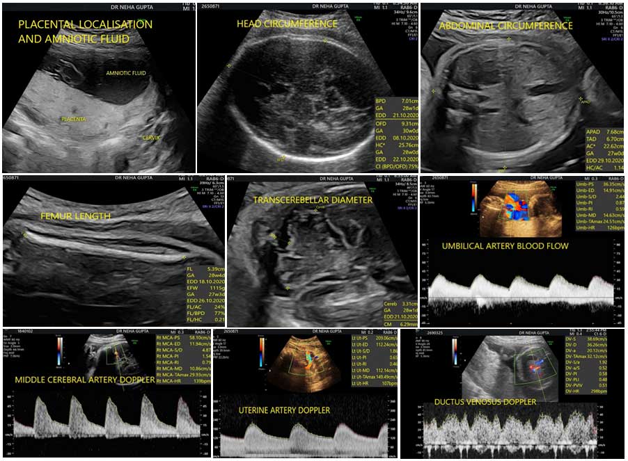
This scan is meant to assess the fetal weight, the fluid around the baby, the placental localisation and the blood flows to the baby.

WHAT DO WE MEAN BY FETAL DOPPLERS?

Fetal dopplers mean assessment of baby’s blood flows. Maternal blood flows to the uterus are assessed by uterine artery Dopplers. Blood flow through cord is called as umbilical artery blood flow. Blood through baby’s brain is called as middle cerebral artery Dopplers and through tummy is referred to as ductus venosus Dopplers.

WHAT IS THE SIGNIFICANCE OF GROWTH AND DOPPLERS SCAN?

The aim is to assess the fetal growth disorders and the well being of the unborn baby. If the growth is appropriate for the weeks, then it is normal. If the growth is small, then it is a weak baby, also called as fetal growth restriction. If the growth is high, then it is a big baby, also called as macrosomia.

Besides fetal weight, maternal history and symptoms, fluid assessment and baby’s blood flows helps us to further identify the babies at risk of adverse outcome. Depending on the above factors, appropriately delivery can be timed.

IS ONE SCAN ENOUGH FOR THE GROWTH ASSESSMENT AND WELL BEING?

No, minimum two growth scans are required to assess the fetal well being. One growth scan gives us the fetal growth assessment at a given time. However, two helps us to establish the growth velocity, and identify the at risk fetuses.

CAN STRUCTURAL ASSESSMENT BE DONE AT THE TIME OF GROWTH SCAN?

A detailed structural assessment is difficult after 26-28 weeks due to fetal position and baby does not move around completely as it does around 20 weeks. But still, whatever is possible due to fetal position and advanced gestational age, is assessed at the time of growth scans.

CAN ANY STRUCTURAL ABNORMALITY BE DETECTED AT THE TIME OF GROWTH SCANS?

Yes, it is possible. The risk of detection of structural abnormality in the third trimester is 1 in 200 pregnancies. These could be those that develop later on like certain brain abnormalities, kidney problems or those which were missed at the time of anomaly scan.

WHEN THE GROWTH AND DOPPLERS SCAN IS DONE?

Usually, the growth and Doppler assessment start from 28 weeks onwards. Most of the times, two scans are done for the growth assessment- one around 28 weeks and other between 34-37 weeks. Sometimes, depending on the growth and blood flows or specific history, more scans may also be required. This will be explained to you by the fetal medicine specialist.

IS 3D/ 4D SCANNING PART OF THIS SCAN?

No. But at this time, if the fetal position is appropriate, good 3D and 4D views can be obtained of the face of the fetus. We routinely take 3D/4D views of the face if the baby’s position is appropriate.

WHO CAN PERFORM GROWTH AND DOPPLERS ASSESSMENT?

Individuals who routinely perform obstetric scans and have the necessary requirements as per the law of the land.

IS PRENATAL ULTRASONOGRAPHY SAFE?

Prenatal ultrasonography appears to be safe for clinical practice. To date, there has been no independently confirmed study to suggest otherwise.

Growth and Dopplers
WHAT IS MULTIPLE PREGNANCY?

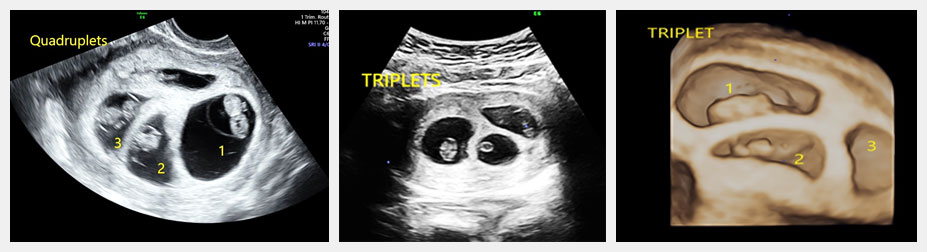
A multiple pregnancy is one in which the mother carries more than one baby in her womb. Most commonly is it two called as twins. If there are three, it is called as triplet and if four, it is quadruplet pregnancy.

WHAT IS THE DIFFERENCE BETWEEN NON-IDENTICAL AND IDENTICAL TWIN/ MULTIPLE PREGNANCY?

Fertilization of egg by a sperm is called zygote which later develops into embryo and then baby.

In non-identical twins (DIZYGOTIC), each egg is fertilized by different sperm. It is like two/ multiple babies with different genetic material growing in the womb at the same time.

This usually happens two or more eggs ovulate at the same time and get fertilized or two or more embryos were transferred in intro-fertilization process.

Identical twins (MONOZYGOTIC) develop from one egg and one sperm forming one zygote. Very early in the process, the fertilized egg or zygote splits into two or more embryos.

This is the concept of ZYGOSITY.

WHAT ARE THE TYPE OF TWIN PREGNANCIES?

Monozygotic are the identical twins. They can be

  • Dichorionic Diamniotic- when each baby has their own placenta and sacs.
  • Monochorionic Diamniotic- When there is single placenta, the placenta is shared among the twins and each twin has their own sac.
  • Monochorionic Monoamniotic- When the twin shares the placenta as well as the sac- single placenta and single sac.

Dizygotic will always be dichorionic and diamniotic- each baby has their own placenta and sacs.

This the concept of CHORIONICITY.

Multiple Pregnancy Scans
WHAT IS CHORIONICITY?

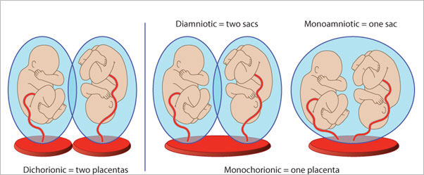
Chorionicity is to determine the type of placentation. Hence, we determine chorionicity and zygosity on the ultrasound.

Determination of chorionicity is important as it determines the type of complications the babies can have and the type of follow up they require.

Multiple Pregnancy Scans
WHAT ARE THE ULTRASOUNDS THAT HAPPEN IN THE MULTIPLE PREGNANCY?
  • Early viability scan- this scan is to determine the number of fetuses and type of multiple pregnancy, that is to determine the CHORIONICITY.
  • First trimester screening- at 11-13+6weeks- Here, we determine the number of fetuses, type of multiple pregnancy, dating, structural abnormalities and screen for chromosomal abnormalities.
  • If it is monochorionic diamniotic pregnancy, then screening for specific complications like twin to twin transfusion syndrome where one fetus bleeds into the other or selective weak baby starts from 16 week onwards. Then the scans are done every 2 weekly till 24 weeks.
  • Anomaly scan -done between 18-20 weeks- to do detailed evaluation of the structural abnormalities of the babies.
  • Growth and Doppler assessment – to assess fetal weight, fluid around the baby and their respective blood flows. It is done every 4 weekly after anomaly scan in dichorionic diamniotic pregnancies and every 2-3 weekly in monochorionic pregnancies.
DOES EVERY MULTIPLE PREGNANCY REQUIRE THE SAME PROTOCOL OF MONITORING?

The care has to be individualised for every multiple pregnancy. As these pregnancies are at risk of early delivery, weak babies, developing high blood pressure and sugars, hence the monitoring of babies by ultrasound needs to be individualised.

Also, the monochorionic pregnancies have unique set of complications like twin to twin transfusion syndrome (TTTS) where one baby bleedss into the other twin or selective fetal growth restriction, where one baby grows really small in comparison to the other baby. Hence, depending on how each multiple pregnancy behaves, we have to alter the protocol.

I can understand that these are somewhat difficult to understand but if you have any queries, I would be eager to answer all of them.

WHAT IS CHORIONIC VILLUS SAMPLING?
Chorionic villus sampling (CVS), or chorionic villus biopsy, is a prenatal test that involves taking a sample of tissue from the placenta to test for chromosomal abnormalities and certain other genetic problems. The placenta is a structure in the uterus that provides blood and nutrients from the mother to the fetus. The chorionic villi are tiny projections of placental tissue that look like fingers and contain the same genetic material as the fetus. It is these villi which are used for genetic testing
WHAT IS THE PURPOSE OF CHORIONIC VILLUS SAMPLING?
Chorionic villus sampling may be used for genetic and chromosome testing in the first trimester of pregnancy. Here are some reasons that a woman might elect to undergo CVS:
Previously Affected Child Or A Family History Of A Genetic Disease, Chromosomal Abnormalities, Or Metabolic DisorderIf An Ultrasound Identifies Structural Abnormality In The Unborn BabyIf Prenatal Screening Shows There Is An Increased Chance Of A Chromosomal Problem Like Down Syndrome,
There may be other reasons for your doctor to recommend a chorionic villus sampling. Getting the test done is completely your choice. We can suggest what is good practice based on evidence-based medicine. Knowing all the pros and cons, you can accept or decline the testing.
WHEN IT IS DONE?
CVS is usually done between 11-12 weeks of pregnancy.
WHAT PRECAUTIONS ARE TAKEN PRIOR TO THE PROCEDURE?
The fetal medicine specialist will explain the procedure to you and offer you the opportunity to ask any questions that you might have about the procedure. You will be asked to sign few consent forms that gives the permission to do the procedure. There is no special restriction on diet or activity prior to chorionic villus sampling. Your doctor will note if you are allergic to any medications, latex, iodine, tape, and anesthetic agents (local and general), medications that you are taking, any history of bleeding disorders or any anticoagulant (blood-thinning) medications, aspirin, or any other medications that may affect blood clotting. It may be necessary for you to stop these medications prior to the procedure. Your blood group will also be noted and if you are Rh negative, an injection anti-D will be given to you after the procedure.
HOW IT IS DONE?
An ultrasound will be performed to check the fetal heart rate and the position of the placenta and the fetus. The fetus is also assessed for structural abnormality. CVS can be done abdominally and transcervically. We generally do it transabdominally. A local anesthetic is injected into the skin. You will feel a needle stick when the anesthetic is injected. This may cause a brief stinging sensation. Then, ultrasound will be used to help guide a long, thin, hollow needle through your abdomen and into the uterus and placenta. This may be slightly painful, and you may feel a cramp as the needle enters the uterus. Tissue will be gently suctioned into a syringe. More than one sample may be needed to obtain enough tissue for testing. The needle will then be removed. An adhesive bandage will be placed over the abdominal needle insertion site.
WHAT HAPPENS AFTER THE PROCEDURE?
You are made to rest for half an hour after the procedure. The fetal heart beat is checked and then you are sent home. Most women have no problems, but you might have a bruise, spotting or cramping. If you do, paracetamol is safe to take. If you have a persistent pain, leaking, bleeding or a high temperature, then you need to go to the hospital.
WHAT ARE THE RISKS INVOLVED?
There is minimal risk of miscarriage about 3: 1000. It does not cause any birth defects or any harm to the mother. The risk of infection is also very low, 1 in 1000.
WHOM TO CONSULT?
You can consult with Dr. Neha Gupta. She is one of the best fetal medicine specialists and is experienced in handling lots of pregnant women to take care of their unborn baby. Contact us to get the appointment and discuss it with her.
WHAT IS PERCUTANEOUS UMBILICAL BLOOD SAMPLING (PUBS)?
Percutaneous umbilical blood sampling — also known as cordocentesis— is a diagnostic prenatal test in which a sample of the baby’s blood is removed from the umbilical cord for testing.
WHAT IS THE PURPOSE OF PUBS OR CORDOCENTESIS?
Cordocentesis is used primarily to detect and treat blood conditions, such as fetal anemia — a low amount of healthy red blood cells in a developing baby or for the diagnosis of fetal infections. Cordocentesis is usually done when a diagnosis can’t be made from amniocentesis, chorionic villus sampling, ultrasound or other methods.
Cordocentesis carries a higher risk of complications to the baby than other procedures do. Rarely, cordocentesis might be used to check fetal chromosomes through chromosome microarray or karyotype analysis. Blood obtained through cordocentesis can also potentially be used for other types of genetic studies. Getting the test done is completely your choice. We can suggest what is good practice based on evidence-based medicine. Knowing all the pros and cons, you can accept or decline the testing.
WHEN IT IS DONE?
Cordocentesis, which is usually done after week 18 of pregnancy, can be used to detect certain genetic disorders, blood conditions and infections.
WHAT PRECAUTIONS ARE TAKEN PRIOR TO THE PROCEDURE?
The fetal medicine specialist will explain the procedure to you and offer you the opportunity to ask any questions that you might have about the procedure. You will be asked to sign few consent forms that gives the permission to do the procedure. There is no special restriction on diet or activity prior to chorionic villus sampling. Your doctor will note if you are allergic to any medications, latex, iodine, tape, and anesthetic agents (local and general), medications that you are taking, any history of bleeding disorders or any anticoagulant (blood-thinning) medications, aspirin, or any other medications that may affect blood clotting. It may be necessary for you to stop these medications prior to the procedure. Your blood group will also be noted and if you are Rh negative, an injection anti-D will be given to you after the procedure.
HOW IT IS DONE?
An ultrasound will be performed to check the fetal heart rate and the position of the placenta and the fetus. The fetus is also assessed for structural abnormality. The procedure is done transabdominally. Guided by ultrasound, the fetal medicine specialist will insert a thin, hollow needle through your abdominal wall into your uterus and into the baby’s cord. A small amount of blood from the vein in the umbilical cord will be withdrawn into a syringe, and the needle will be removed. An adhesive bandage will be placed over the abdominal needle insertion site. You’ll need to lie still while the needle is inserted and the blood is withdrawn. You might notice a stinging sensation when the needle enters your skin, and you might feel cramping when the needle enters your uterus.
WHAT HAPPENS AFTER THE PROCEDURE?
You are made to rest for half an hour after the procedure. The fetal heart beat is checked and then you are sent home. Most women have no problems, but you might have a bruise, spotting or cramping. If you do, paracetamol is safe to take. If you have a persistent pain, leaking, bleeding or a high temperature, then you need to go to the hospital.
WHAT ARE THE RISKS INVOLVED?
However, besides, these there is increased risk of bleeding from the cord puncture site or formation of hematoma at the puncture site. There is possibility of slowing of fetal heart rate and fetal loss rate is 1-2%. There is minimal risk of miscarriage or infection after the procedure.
WHOM TO CONSULT?
You can consult with Neha Gupta. She is one of the best fetal medicine specialists and is experienced in handling lots of pregnant women to take care of their unborn baby. Contact us to get the appointment and discuss it with her.

Genetic counseling is the information provided by the counselor as to how certain genetic disease might affect you and your family and what is the risk of transmission to your children. The doctor will first collect your and family history.

It is done in those cases-

If There Is Family History Of Hereditary Disease In Your FamilyIf There Is History Of Affection Of Birth Defect Or Genetic Disease In The Previous ChildIf There Is History Of Certain Findings On Ultrasound In The Present Pregnancy Which Can Point Towards A Genetic Disorder.Abnormal Screening Results Or Invasive Testing Results

After counseling, a repeat scan or certain tests might be prescribed.

WHAT IS 3D/4D ULTRASOUND?
There are 2D, 3D and 4D ultrasounds. All ultrasound uses sound waves to create an image of the unborn baby on the screen. A 2D ultrasound of a fetus creates a black-and-white image. The main scan is done in 2D only. We take images of the unborn baby in grey scale. We take measurements of the unborn baby, assess its growth and assess the structure of the baby mainly in 2D.Nowadays, 3D- three dimensional and 4D – fourth dimensional ultrasounds have become common. Usually, all ultrasound machines have software and volume probes with which we do 3D/ 4D scan.3D scan makes three dimensional images of the unborn baby like that of a face. When we see these images in real time, that is, we see the movements of the unborn baby as well, it is called 4D ultrasound. Some centres have started adding 5D ultrasound also. 5D ultrasound is nothing but an extension of 3D ultrasound only.

WHY ARE 3D AND 4D ULTRASOUND SCANS DONE DURING PREGNANCY?

The main scan is done in grey scale, that is 2D scanning. 3D and 4D ultrasounds can be used by the person to add more information to it but are not absolutely necessary.

What laymen understand is that , by means of 3D and 4D ultrasound , the parents can see realistic images of the face of the baby. It adds to great experience in pregnancy. But it is not limited to taking pictures of baby’s faces.

The benefits of 3D and 4D ultrasound are limited. It becomes useful when any structural abnormality is suspected. It helps us to take views of the baby from different angles and provides more depth to our understanding of abnormalities. 3D and 4D ultrasounds can be done to add more details which helps us in counselling couples. Like when we see a gap in the lips- cleft lip, 3d and 4D ultrasound helps to know the extent of the gap, whether there is involvement of palate also.

3D and 4D ultrasounds are not generally part of daily prenatal tests. What is interesting is that 3D ultrasounds provide the three-dimensional image of your infant, while 4D ultrasounds provide a live visual effect, such as a clip, you can see your baby smile or yawn.